Cardiodynamics & Intelligence

AI that reads the angiogram for blood flow.

Our algorithms segment the coronary tree on every frame, track how contrast moves through it, and detect the disturbed and retrograde flow that injures the vessel wall and sets off coronary artery disease.

Built in Chicago on nearly three decades of clinical research in dynamic angiography, computational fluid dynamics and image segmentation. Founded by Thach N. Nguyen, MD.

About us

Who we are

CardioDynamics and Intelligence is a medical technology company based in Chicago. It brings together interventional cardiologists, physician-scientists and engineers around one question: how does the movement of blood shape heart disease, and how can we measure it well enough to act on it?

The company grew out of nearly three decades of clinical research and teaching in interventional cardiology, and a long-running program on coronary flow built on hydraulics, acoustics and frame-by-frame angiography. We are turning that program into AI algorithms and medical devices.

Founder & CEO

John M. Le, MD, PhD

Physician-scientist

Dr. Le works on large language models, computer vision, machine learning and deep learning applied to cardiology and multimodal imaging — ECG, echocardiography, CT, MRI and digital subtraction angiography — with a focus on clinically deployable AI systems for diagnosis, risk stratification and clinical decision support.

He has led collaborative AI projects with researchers at Harvard Medical School, Johns Hopkins University and Carnegie Mellon University on real-time imaging interpretation and cardiovascular phenotyping. His work has appeared in The Lancet, NEJM, JAMA, EClinicalMedicine, Briefings in Bioinformatics and the IEEE Journal of Biomedical and Health Informatics, with presentations at AHA Scientific Sessions, ACC's Annual Scientific Session, IEEE EMBS, ISBI, ICCV and MICCAI.

Manager · Management Committee

Loc Vu, MD, FACC, FAHA

Cardiologist and Clinical Lecturer, Vietnam

Dr. Vu's research focuses on coronary artery disease — coronary physiology, flow dynamics, and artificial intelligence in cardiovascular imaging. He collaborates on the Global Burden of Disease study at the University of Washington Institute for Health Metrics and Evaluation, and reviews abstracts for the American College of Cardiology Quality Summit and the American Heart Association's Cardiomyopathies Peer Review Committee.

He completed the Global Clinical Scholars Research Training certificate program at Harvard Medical School (2025) and received the Paul Dudley White International Scholar Award at the 2023 AHA Scientific Sessions for the highest-scoring abstract from Vietnam. He serves on the Communications Committee of the AHA's Council on Clinical Cardiology, is a Social Media Ambassador for the Cardiovascular Research Foundation in Seoul, and is a participant in the 2026 cohort of the International Atherosclerosis Society's Inflammation Academy.

"The program gave me far more than just research skills," he says of his time at Harvard. "It changed the way I ask scientific questions, collaborate across cultures, communicate evidence, and think about leadership."

Thach N. Nguyen, MD
Scientific Advisor

Thach N. Nguyen, MD

Interventional cardiologist · FACC, FSCAI

Dr. Nguyen has spent nearly three decades teaching and studying interventional cardiology. He is lead editor of the Practical Handbook of Advanced Interventional Cardiology: Tips and Tricks, now in its 6th edition (Wiley, 2026), and of Management of Complex Cardiovascular Problems, and has authored more than 180 indexed publications since 1998.

He served on the Board of Trustees of the Society for Cardiovascular Angiography and Interventions (SCAI, 2013–2016), has helped organize the Great Wall International Congress of Cardiology since 1992, and is an honorary professor of medicine at Hanoi Medical University. He originated dynamic coronary angiography and the coronary acoustic map.

Prof. C. Michael Gibson
Senior Advisor

Prof. C. Michael Gibson

Professor of Medicine, Harvard Medical School · MS, MD

Prof. Gibson is an interventional cardiologist at Beth Israel Deaconess Medical Center and chief executive of the Baim Institute for Clinical Research and the PERFUSE Study Group, which he founded in 1987. He invented the TIMI frame count and the TIMI myocardial perfusion grade, angiographic measures of coronary blood flow now used worldwide.

The institutes he leads have run more than 1,000 studies across a network of thousands of sites, and he has repeatedly been named among the most highly cited researchers in science.
Scientific advisory board

Kwan S. Lee, MB BCh, MD

Interventional cardiologist, Mayo Clinic Arizona

Product

What we build

AI that turns a routine coronary angiogram into an objective, frame-by-frame record of how blood moves. Everything here is in development and under clinical study.

01

Flow-detection AI

Algorithms that segment the coronary tree, detect retrograde flow and track contrast density frame by frame on the angiogram.

02

Medical devices

Devices in development that pair our algorithms with angiographic imaging to detect and measure blood flow at the point of care.

03

CFD modeling

Patient-specific models of shear stress, recirculating flow and cavitation in bifurcation and left main disease.

Flow explorer

What we see in the coronary arteries

Drag the slider through the cardiac cycle and tap the numbered points on each artery. The animations are schematic; the numbers come from our published angiographic series.

Diastole Systole
→←

01 · Healthy pattern

Laminar flow

Blood moves in smooth, parallel layers: fastest at the center, slowest at the wall. This is healthy flow, and it was the dominant pattern in our series.

Fluids, 2024 · n = 51
55%of right coronary arteries showed laminar flow
Tap a numbered point on the artery to see what happens there.
Diastole Systole
→←

02 · Disturbed pattern

Retrograde flow

“Retrograde” means backward. For most of the beat all the blood moves forward (blue). At the switch from diastole to systole, the layers next to the wall stop and run backward (red) while the center still moves forward. In life this lasts only a few frames, so the animation replays that moment in slow motion.

Fluids, 2024 · n = 51
8%of right coronary arteries, every case with systolic pressure above 120 mmHg
Tap a numbered point on the artery to see what happens there.
Diastole Systole
→←

03 · Where lesions sit

Collision line

Antegrade and retrograde streams meet head-on. The pressure and turbulence at that line injure the wall.

Eur J Anat, 2021
75%of mild-to-moderate lesions sat exactly at the collision line
Tap a numbered point on the artery to see what happens there.
Diastole Systole
● rarefaction● compression

04 · Acoustic theory

Water hammer

When flow is checked abruptly, a pressure wave runs back up the artery and reflects at branches and narrowings. Forward and reflected waves add up to a standing wave along the vessel.

Diagnostics, 2025
A · Ncompression antinodes lined up with severe stenoses; nodes stayed free of lesions
Tap a numbered point on the artery to see what happens there.

Methods

How we study it

Acoustics, computational fluid dynamics and image segmentation, each shown here as a schematic.

Acoustic theory of blood flow

When flow is checked abruptly at the switch from diastole to systole, a pressure wave runs back up the artery: the water hammer. It reflects at branches and narrowings, and the forward and reflected waves (dashed) add up to a standing wave. Compression antinodes (A) lined up with severe stenoses; nodes (N) stayed free of lesions.

Diagnostics, 2025

Computational fluid dynamics

Patient-specific models show a fast jet through the narrowing and slow, recirculating flow just beyond it, where shear stress on the wall drops. In left main stenoses averaging 72%, the models produced vapor bubbles whose collapse reached peak pressures of 3.9 MPa before the carina.

Rev Cardiovasc Med, 2022 · n = 12

Coronary segmentation

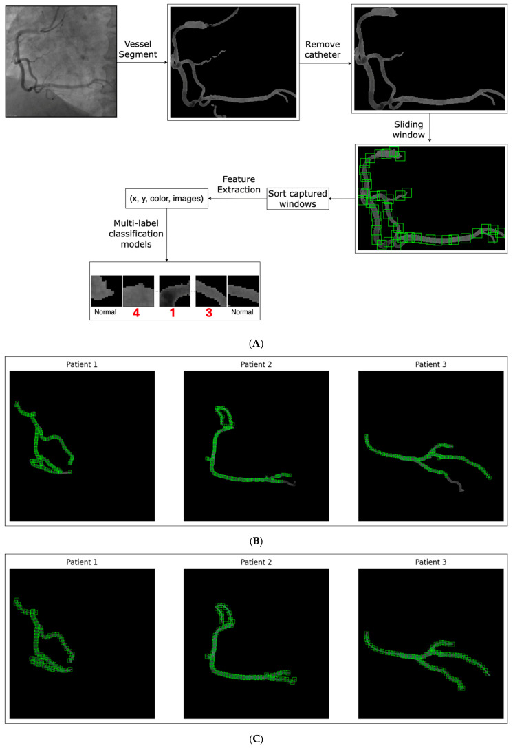
A deep-learning model traces the coronary tree on every angiographic frame and follows the centerline of each vessel. Tracking how contrast density changes along those centerlines, frame by frame, turns the angiogram into a measurable record of flow.

In development
diastoleDrag to rotate · + − to zoom

3D reconstruction: CFD, retrograde flow and water hammer

Once the tree is segmented, its centerlines can be rebuilt as a 3D model and the blood inside it simulated. Here the particles are blood: they speed up through the narrowing on the LAD (red), and just beyond it slow, reversed flow hugs the wall, where shear stress drops (blue wall). Those are the zones the CFD models single out.

Schematic · in development

From our angiograms

Flow, seen frame by frame

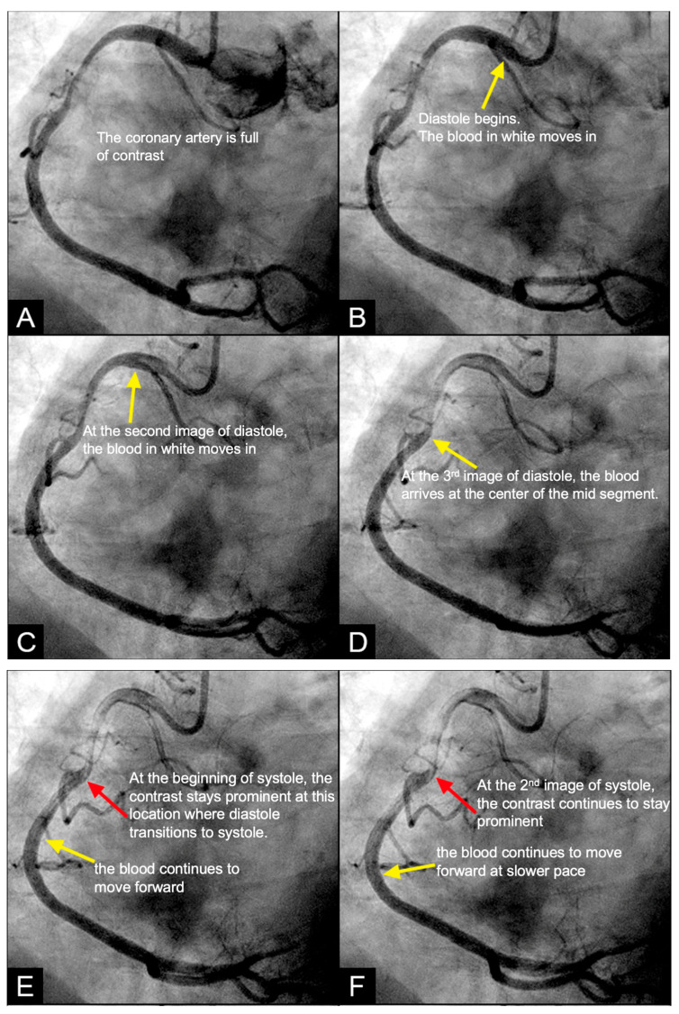
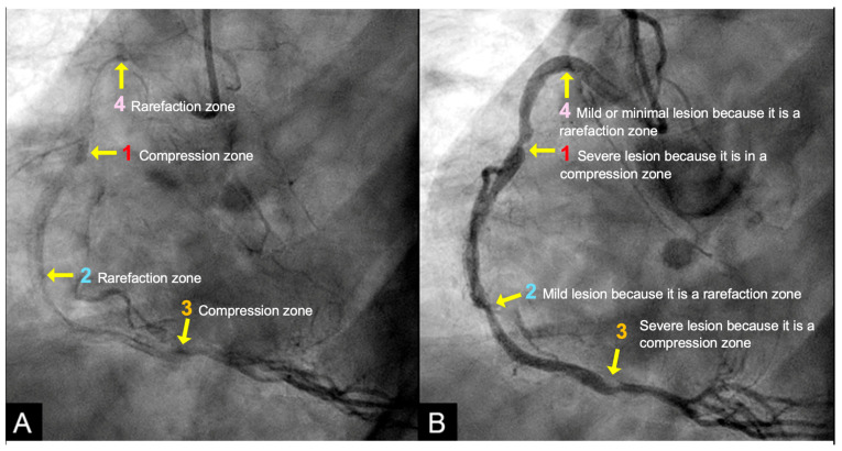
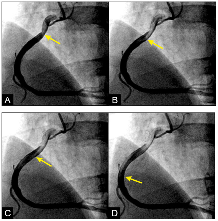
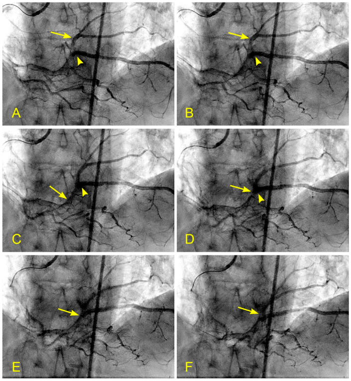
Selected figures from our published studies. Each series was filmed at 15 frames per second; in these images blood appears white and contrast black.

Four consecutive angiographic frames of a right coronary artery

Laminar flow

Blood (white) enters the right coronary artery with a sharp, pointed front, pushing contrast (black) ahead of it in organized layers.

Diagnostics 2025;15(5):553, Figure 3 · CC BY 4.0
Six sequential angiographic frames of a distal right coronary artery

Reverse flow

Blood moves forward past the origin of the posterior descending artery, then contrast is seen travelling back up the distal right coronary artery.

Diagnostics 2024;14(12):1282, Figure 15 · CC BY 4.0
Consecutive angiographic frames of a right coronary artery 67 milliseconds apart

Collision

In frames 67 ms apart, antegrade and retrograde streams meet in the right coronary artery at the site of a moderate mid-segment lesion.

Diagnostics 2025;15(5):553, Figure 7 · CC BY 4.0
End-diastolic angiogram of a right coronary artery with residual pockets of contrast

Contrast pockets

At end-diastole most contrast has washed out, but dense pockets persist: stagnation zones that may mark compression and rarefaction zones of a standing pressure wave.

Diagnostics 2025;15(5):553, Figure 15 · CC BY 4.0
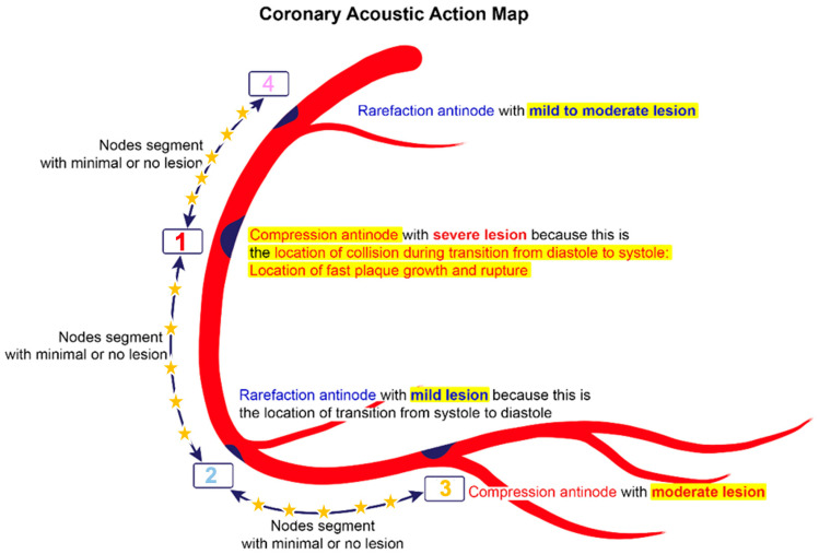
Diagram of a coronary acoustic action map drawn on a right coronary artery

Coronary acoustic action map

Severe lesions sit at compression antinodes, milder lesions at rarefaction antinodes, and the node segments between them show minimal or no disease.

Diagnostics 2025;15(5):553, Figure 19 · CC BY 4.0
AI segmentation of coronary arteries and catheter at three window sizes

AI segmentation

An AI algorithm segments the coronary arteries and the catheter on the angiogram, shown at window sizes of 10, 15 and 20 pixels.

Diagnostics 2025;15(5):553, Figure 5 · CC BY 4.0

Figures reproduced without modification from open-access articles in Diagnostics (MDPI), published under the Creative Commons Attribution 4.0 license.

Flagship program

The coronary acoustic map

Why do lesions form where they do? Dr. Nguyen’s long-running program reads the angiogram frame by frame for flow, borrowing from hydraulic engineering: flow collision, cavitation and water hammer. Reflected pressure waves form nodes and antinodes along the artery — and the hypothesis is that lesions gather at the antinodes.

● pressure rise● pressure drop

Water hammer in everyday life

Shut a tap quickly and the pipes bang. The moving water has nowhere to go, so its momentum turns into a pressure wave that races back up the pipe, reflects at the far end and shakes the pipe until it dies away. Engineers call it water hammer, and it can crack pipes. Our program asks whether the same thing happens in a coronary artery each time forward flow is checked at the switch from diastole to systole.

Everyday analogy · schematic
A antinodeN node

Standing waves in everyday life

Pluck a guitar string and a wave runs to each end, reflects and comes back. The two waves, travelling in opposite directions, add up to a pattern that stays in place: some points never move (nodes, N) and the points between them swing the hardest (antinodes, A). Touch the string lightly at the right spot and you force more loops, which is how a guitarist plays a harmonic. The acoustic map looks for the same pattern in the pressure inside an artery.

Everyday analogy · schematic
A antinodeN node
Aim 1 · Identify

Codify existing lesions

Locate compression and rarefaction zones on the angiogram and relate them to lesions already present.

Aim 2 · Forecast

Predict progression

Use serial angiograms to test whether lesions at antinodes grow faster than lesions elsewhere.

Aim 3 · Predict

Locate future lesions

Forecast where new disease will appear on segments that look normal today.

Our hypothesis

Retrograde flow injures the artery

Dynamic angiography lets us watch contrast move frame by frame, and it shows that blood does not always travel forward. At the transition from diastole to systole, antegrade and retrograde streams can meet head-on — the collision line — and reflected pressure waves travel back up the artery.

75%
of mild-to-moderate lesions sat at the collision line, where antegrade and retrograde flow meet Eur J Anat, 2021
47%
of patients showed retrograde flow in the iliac artery; 75% of them had diastolic pressure above 80 mmHg (p < 0.001) Fluids, 2024 · n = 51
8%
showed retrograde flow in the right coronary artery, all with systolic pressure above 120 mmHg; an AI algorithm was trained to detect it Fluids, 2024 · n = 51

Clinical trials

Trials & clinical studies

From first-in-man device studies to multicenter cohorts, our team has designed, enrolled and published prospective clinical research across three continents.

StudyDesignStatusPublication
PADN-1 First-in-man pulmonary artery denervation for pulmonary arterial hypertension · prospective, single-center Published JACC 2013 · DOI
VSTENT Bioresorbable-polymer sirolimus-eluting stent in de novo coronary lesions · prospective, multicenter cohort Published Cardiovasc Diagn Ther 2023 · DOI
Robotic PCI Safety and feasibility during early implementation of robotic-assisted PCI Published Front Cardiovasc Med 2026 · DOI
TAVR ≥80 vs <80 Transcatheter aortic valve replacement at moderate surgical risk · observational, Vietnam Published Cardiovasc Innov Appl 2024 · DOI
Coronary Acoustic Map Diagnostic and predictive accuracy of AI-read dynamic angiography · longitudinal cohort Protocol published Diagnostics 2025 · DOI
For sponsors & CROs
  • Investigator-initiated trial design and protocol writing
  • Site identification and feasibility across our network
  • Angiographic core-lab reading, including dynamic angiography
  • AI-assisted image analysis, coronary segmentation and CFD modeling
  • Statistical analysis, meta-analysis and manuscript preparation
Sites & network
  • Collaborating centers in the United States, China, Italy, Vietnam, Bulgaria and Poland
  • Catheterization laboratories with high-volume coronary and structural programs
  • Active trials and participating sites are listed here as agreements are finalized

Vision

Every angiogram read for flow, not only for narrowing

Today an angiogram tells a physician where an artery is narrowed. We are building the AI algorithms and medical devices that detect blood flow itself, so the same study can explain why a lesion formed there and show where the next one is likely to appear.

Understand

Explain the disease

Establish how disturbed and retrograde flow injures the coronary wall, with studies that others can repeat.

Build

Detect flow with AI

Build algorithms that turn routine angiograms into objective, automated measurements of blood flow.

Deliver

Put it in clinicians’ hands

Develop and validate medical devices that bring flow detection into the catheterization laboratory.

Research

Research areas

Four areas, all aimed at detecting and measuring coronary blood flow.

Flow & imaging

Dynamic angiography

Frame-by-frame reading of coronary flow: antegrade and retrograde streams, the collision line and reflected pressure waves.

Artificial intelligence

AI & machine learning

Algorithms that detect retrograde flow on the angiogram and protocols that predict where coronary lesions will form.

Computational modeling

Computational fluid dynamics

Patient-specific models of bifurcation and left main stenting, shear stress, recirculating flow and cavitation.

Computer vision

Coronary segmentation

Automated, frame-by-frame segmentation of the coronary tree and tracking of contrast density across the cardiac cycle.

Publications

Selected work

A cross-section of our team’s clinical and translational research. Full list via Scopus Author ID 7404371747.

Coronary flow and interventional cardiology

  1. 2026

    Practical Handbook of Advanced Interventional Cardiology: Tips and Tricks, 6th edition

    John Wiley & Sons · edited by Thach N. Nguyen and colleagues

  2. 2026

    Safety and Feasibility During Early Implementation of Robotic-Assisted Percutaneous Coronary Intervention

    Frontiers in Cardiovascular Medicine 13:1731900 · 10.3389/fcvm.2026.1731900

  3. 2025

    Water Hammer Phenomenon in Coronary Arteries: Scientific Basis for Diagnostic and Predictive Modeling with Acoustic Action Mapping

    Diagnostics 15(5):553 · 10.3390/diagnostics15050553

  4. 2024

    Transcatheter Aortic Valve Replacement in Patients ≥80 and <80 Years of Age with Aortic Valve Stenosis at Moderate Surgical Risk

    Cardiovascular Innovations and Applications 9:1–11 · 10.15212/CVIA.2024.0003

  5. 2023

    Trends in Cardiogenic Shock–Related Mortality in Patients With Acute Myocardial Infarction in the United States, 1999 to 2019

    American Journal of Cardiology 200:18–25 · 10.1016/j.amjcard.2023.05.026

  6. 2023

    Safe and Effective Profile of the VSTENT Bioresorbable Polymer Sirolimus-Eluting Stent in De Novo Coronary Artery Lesions: A Prospective, Cohort, Multicenter Study

    Cardiovascular Diagnosis and Therapy 13(3):474–486 · 10.21037/cdt-22-522

  7. 2022

    Distal Snuffbox Versus Conventional Radial Artery Access: An Updated Systematic Review and Meta-Analysis

    Journal of Vascular Access 23(4):653–659 · 10.1177/11297298211005256

  8. 2020

    Impella RP in Hemodynamically Unstable Patients with Acute Pulmonary Embolism

    Journal of Artificial Organs 23(2):105–112 · 10.1007/s10047-019-01149-9

  9. 2018

    Complex Coronary Bifurcation Treatment by a Novel Stenting Technique: Bench Test, Fluid Dynamic Study and Clinical Outcomes

    Catheterization and Cardiovascular Interventions 92(5):907–914 · 10.1002/ccd.27494

  10. 2017

    Microbiological Profile and Risk Factors for In-Hospital Mortality of Infective Endocarditis in Tertiary Care Hospitals of South Vietnam

    PLoS ONE 12(12):e0189421 · 10.1371/journal.pone.0189421

  11. 2013

    Pulmonary Artery Denervation to Treat Pulmonary Arterial Hypertension: The Single-Center, Prospective, First-in-Man PADN-1 Study

    Journal of the American College of Cardiology 62(12):1092–1100 · 10.1016/j.jacc.2013.05.075

Cardiovascular AI

  1. 2026

    Imaging-Anchored Multiomics in Cardiovascular Disease

    Briefings in Bioinformatics 27(4):bbag365 · PMC13418848

  2. 2026

    Protective Predictors of Cardiovascular Disease: An Explainable AI Approach

    Public Health 250:106050 · 10.1016/j.puhe.2025.106050

  3. 2026

    Ultra-ECP: Ellipse-Constrained and Point-Robust Foundation Model Adaptation for Fetal Cardiac Ultrasound Segmentation

    Medical Imaging with Deep Learning (MIDL) 2026 · Proceedings

  4. 2026

    A Real-Time Uncertainty-Aware Digital Twin Framework for Intra-Operative Valve Surgery

    Annual Reliability and Maintainability Symposium (RAMS) 2026 · 10.1109/RAMS50514.2026.11424435

  5. 2025

    3-D TEE Mitral Valve Segmentation and Mesh Reconstruction with Real-Time Quality Assurance

    IEEE-EMBS International Conference on Biomedical and Health Informatics (BHI) 2025 · 10.1109/BHI67747.2025.11269550

  6. 2025

    Integrating Advanced Sensing Technologies and Artificial Intelligence for Predicting Cardiovascular Risks

    Book chapter, Cutting-Edge Diagnostic Technologies in Cardiovascular Diseases · 10.1201/9781003481621-11

  7. 2025

    OASIS-Net: An Obstetric Adversarial Semi-Supervised Image Segmentation Network for Ultrasound Imaging

    IEEE Journal of Biomedical and Health Informatics · 10.1109/JBHI.2025.3631102

Presentations

  1. 2025

    Cardiac Involvement in Hepatocellular Carcinoma: A Systematic Review and Meta-Analysis

    AHA Basic Cardiovascular Sciences (BCVS) 2025, Baltimore · Circulation Research 137(Suppl 1) · 10.1161/res.137.suppl_1.Fri073

  2. 2023

    Successful Treatment of Amniotic Fluid Embolism Complicated by Multiple Cardiac Arrest with VA-ECMO

    American College of Cardiology Annual Scientific Session (ACC) 2023

Patents

Intellectual property

Our algorithms and devices are in development. We are evaluating intellectual property protection for them. For licensing and partnership inquiries, contact us.

Partner with us

Work with us

We work with medical device and pharmaceutical companies, contract research organizations, hospitals and clinical sites that need rigorous answers about coronary flow and interventional cardiology.

Contact us

  • Sponsors & CROsDevice and drug studies, trial design and angiographic core-lab analysis.
  • Hospitals & clinical sitesEnrollment in multicenter studies and registries.
  • AI & imagingCoronary segmentation, CFD modeling and predictive analytics as a service.
  • Development partnersJoint development and validation of flow-based diagnostic software.

Contact

Get in touch

For partnerships, clinical studies, core-lab and AI services, or general questions.

Email
contact@cardiodynamicsintelligence.com
Location
Chicago, Illinois, USA